CSCT / CTI – Advanced Materials for Biomedical Applications
Advanced Materials for Biomedical Applications – CSCT / CTI Seminar
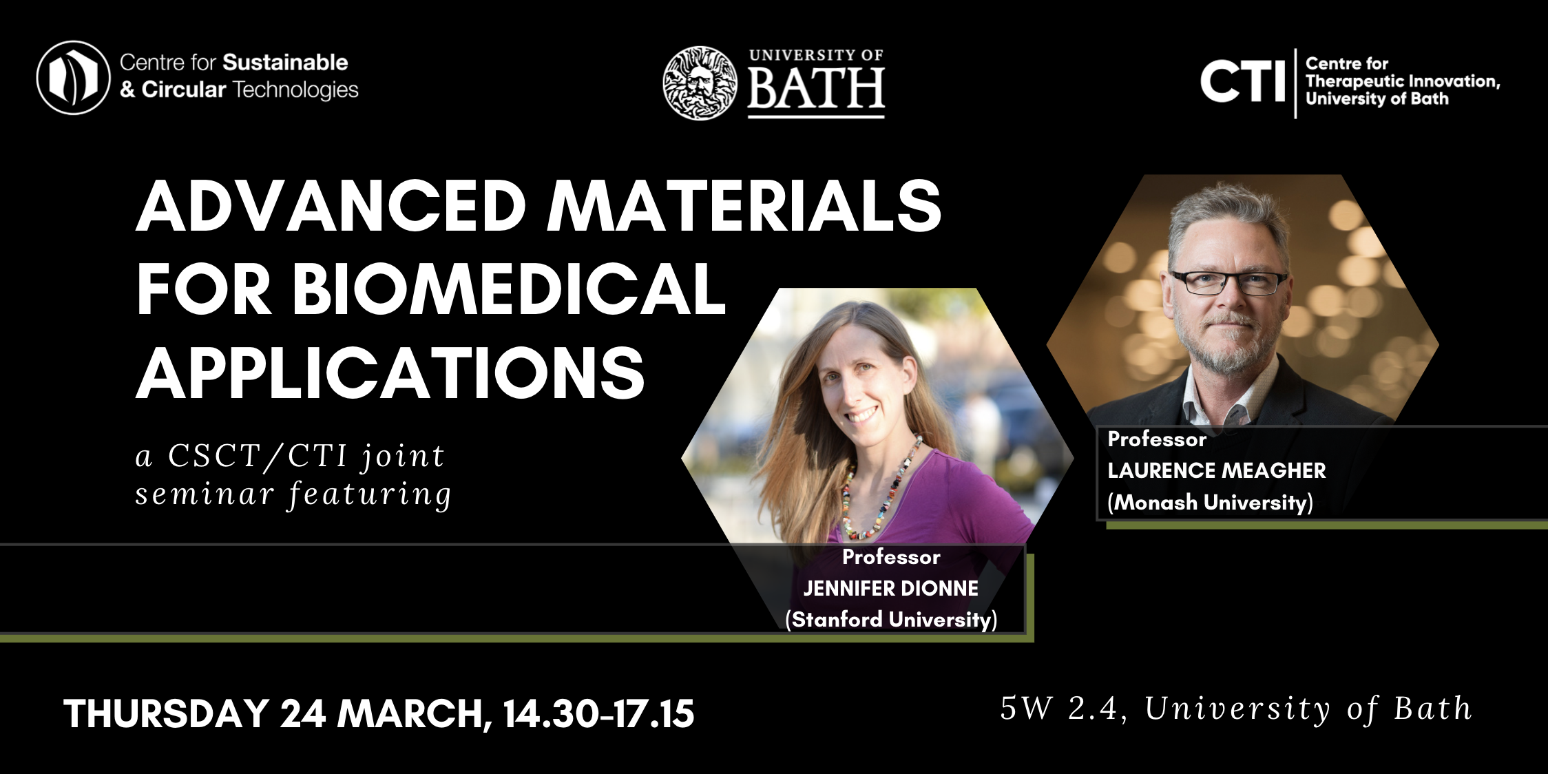
The Advanced Materials for Biomedical Applications jointly sponsored by the Centre for Sustainable & Circular Technologies and the Centre for Therapeutic Innovation. We are delighted to host talks from Professor Laurence Meagher at Monash University and Professor Jen Dionne at Stanford University. The seminar will take place on Thursday 24th March, from 14:30-17:15, in 5 West 2.4.
The confirmed speakers are:
- Jessica Lucena-Thomas, CSCT Alumna: Manufacturing patient derived organoids for use in drug discovery and development
- Prof Laurence Meagher: Electrospinning and tissue bioprinting for the generation of 3D cell culture microenvironments
- Professor Jen Dionne: Emerging nanophotonic platforms for infectious disease diagnostics: Re-imagining the conventional microbiology toolkit
Please note, this is an in-person seminar, with Prof Dionne’s seminar broadcast at the lecture theatre. Prof Dionne’s talk will be recorded and available on the CTI website. Talk abstracts and biographies of the key speakers are below.
Speakers
Professor Laurence Meagher, Monash University
Director, ARC Training Centre for Cell and Tissue Engineering Technologies
Director, SPARK Monash Program
Talk title: Electrospinning and tissue bioprinting for the generation of 3D cell culture microenvironments
ABSTRACT
In this talk I will discuss the culture of cells in 3D matrices in two very different microenvironments. The first microenvironment is prepared using a technique called electrospinning which produces fibrous matts where the individual fibres have sub-micron diameters. The fibres, which are degradable, have been surface modified with peptide decorated, synthetic polymer brush coatings. This produces a 3D microenvironment which supports cell attachment and growth where the mechanical properties of the fibres are modified by the coating, which engages very specifically with cells and has very low non-specific biomolecule adsorption. An example includes use with mesenchymal stromal cells.
The second microenvironment is that of 3D bioprinted constructs where the cell carrier materials are self-assembling hydrogel systems which are printed using extrusion techniques. In this part of the talk, I will briefly discuss three different systems for three very different tissue engineering applications (cardiac, neural and vascular) and highlight some of the interesting features of these type of systems, key features of bioink carrier materials as well discuss some of the challenges faced by the field.
BIOGRAPHY
Laurence Meagher is the Director of the ARC Training Centre in Cell and Tissue Engineering Technologies (https://ctet.org.au/), Professor in Monash University’s Department of Material Science and Engineering and Director of the SPARK program at Monash University. He has worked widely in the field of biomedical materials and devices since 1999. Prior to joining Monash University, he worked at CSIRO Manufacturing. Over the last 20 years or so his research focus has been in the development and translation of biomedical materials and more specifically in the biomaterials and biointerfaces area. He has participated in and led programs leading to marketed products, most notably in the area of silicone hydrogel contact lenses, where he was part of a team that developed the CIBA Vision’s second-generation Air OptixTMproduct. Over the last decade, his research has largely been focused on the development of bioactive surface coatings for commercial application, for example, in the development of materials for ex vivo stem cell expansion in the CRC for Polymers, vehicles for drug delivery, “stealth” and bioactive coatings for biomedical implants, antibacterial materials and surface coatings for biomedical devices and most recently implantable medical devices.
Professor Jen Dionne, Stanford University
Senior Associate Vice Provost of Research Platforms/Shared Facilities
Associate professor of Materials Science and Engineering and, by courtesy, of Radiology
Talk title: Emerging nanophotonic platforms for infectious disease diagnostics: Re-imagining the conventional microbiology toolkit
ABSTRACT
We present our research controlling light at the nanoscale for infectious disease diagnostics, including detecting bacteria at low concentration, sensing COVID gene sequences, and visualizing in-vivo inter-cellular forces. First, we combine Raman spectroscopy and deep learning to accurately classify bacteria by both species and antibiotic resistance in a single step. We design a convolutional neural network (CNN) for spectral data and train it to identify 30 of the most common bacterial strains from single-cell Raman spectra, achieving antibiotic treatment identification accuracies exceeding 99% and species identification accuracies similar to leading mass spectrometry identification techniques. Our combined Raman-CNN system represents a proof-of-concept for rapid, culture-free identification of bacterial isolates and antibiotic resistance. Second, we describe resonant nanophotonic surfaces, known as “metasurfaces” that enable multiplexed detection of SARS-CoV-2 gene sequences. Our metasurfaces utilize guided mode resonances excited in high refractive index nanostructures. The high quality factor modes produce a large amplification of the electromagnetic field near the nanostructures that increase the response to targeted binding of nucleic acids; simultaneously, the optical signal is beam-steered for multiplexed detection. We describe how this platform can be manufactured at scale for portable, low-cost assays. Finally, we introduce a new class of in vivo optical probes to monitor biological forces with high spatial resolution. Our design is based on upconverting nanoparticles that, when excited in the near-infrared, emit light of a different color and intensity in response to nano-to-microNewton forces. The nanoparticles are sub-30nm in size, do not bleach or photoblink, and can enable deep tissue imaging with minimal tissue autofluorescence. We present the design, synthesis, and characterization of these nanoparticles both in vitro and in vivo, focusing on the forces generated by the roundworm C. elegans as it feeds and digests its bacterial food.
BIOGRAPHY
Jennifer Dionne is the Senior Associate Vice Provost of Research Platforms/Shared Facilities and an associate professor of Materials Science and Engineering and, by courtesy, of Radiology at Stanford. She is also an Associate Editor of Nano Letters, director of the DOE-funded Photonics at Thermodynamic Limits Energy Frontier Research Center, and an affiliate faculty of the Wu Tsai Neurosciences Institute, the Institute for Immunity, Transplantation, and Infection, and Bio-X. Jen received her B.S. degrees in Physics and Systems Science and Mathematics from Washington University in St. Louis, her Ph. D. in Applied Physics at the California Institute of Technology in 2009, and her postdoctoral training in Chemistry at Berkeley. Her research develops nanophotonic methods to observe and control chemical and biological processes as they unfold with nanometer scale resolution, emphasizing critical challenges in global health and sustainability. Her work has been recognized with the Alan T. Waterman Award, a NIH Director’s New Innovator Award, a Moore Inventor Fellowship, the Materials Research Society Young Investigator Award, and the Presidential Early Career Award for Scientists and Engineers, and was featured on Oprah’s list of “50 Things that will make you say ‘Wow’!”. Beyond the lab, Jen enjoys exploring the intersection of art and science, long-distance cycling, and reliving her childhood with her two young sons.